Diatom frustules: nature's microscopic glass architecture
Imagine a single-celled organism making its own intricately perforated glass shell. Diatoms have been building such structures in oceans, lakes and waterways for millions of years. This interactive visualisation invites you to inspect one of those architectural ideas in three dimensions.
What is a diatom?
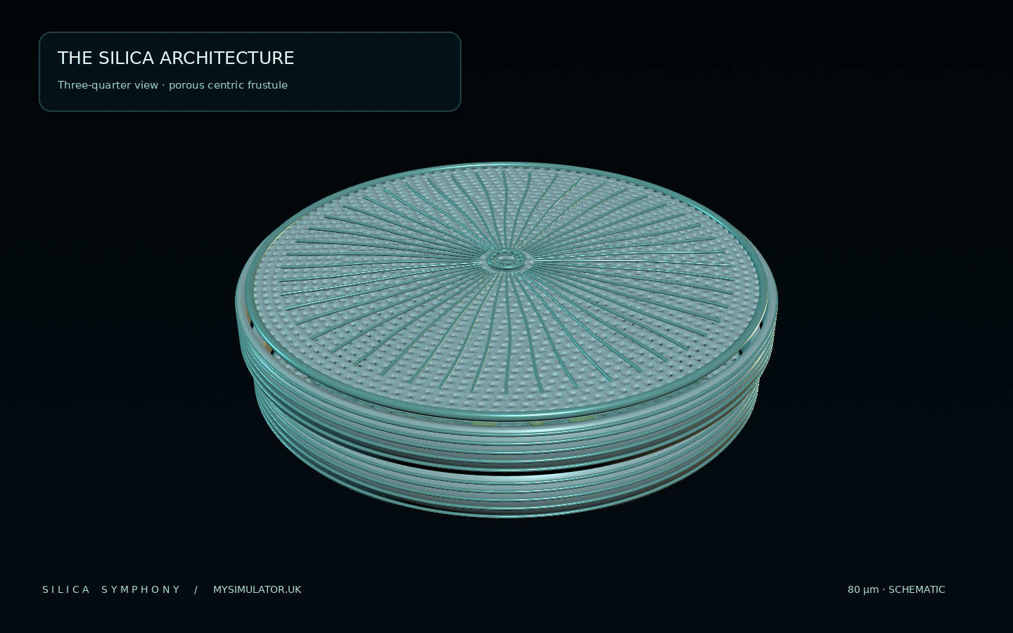
A diatom is a microscopic photosynthetic organism found in a wide variety of aquatic environments. These single-celled eukaryotes are well known for their mineralised cell walls made predominantly of biogenic, hydrated, amorphous silica. Their external architecture can show strikingly regular patterns when viewed with a microscope. Different taxa vary widely in size, shape, symmetry, pore design and way of life; an elongated pennate diatom will not look like a radially patterned centric one.
Diatoms are part of the phytoplankton that support aquatic food webs. For scientists, their distinctive silica parts also provide features useful for biological identification, palaeoenvironmental work and research on microstructured materials. The 3D model on this page is a new, fictional centric-type illustration inspired by well-documented shell design principles, rather than a digitally scanned or identified specimen.
Two parts of the silica frustule
A diatom's mineral shell is called a frustule. In a common architectural arrangement it consists of two overlapping halves, the epitheca and hypotheca. The upper half is the slightly larger one. Each half includes a larger valve face and an adjoining side assembly called a girdle, or cingulum. The arrangement is often compared to a shallow box with a lid: one half can fit inside the other.
Epitheca
Typically the larger half. Its valve face can carry fine patterns of pores, thickened ribs and taxonomically useful features.
Hypotheca
The smaller matching half. In our model it has its own separately constructed porous valve surface and visualised support ribs.
Cingulum
The overlapping girdle bands make the sidewall more than a single plain cylinder. Bands can be added during cell growth.
Why are the valve faces full of holes?
Microscopic openings in a diatom valve are commonly called areolae. In centric diatoms the perforations often participate in radially ordered patterns. Their exact shapes and arrangements depend on the species, and an areola may be much more complicated than a simple hole: internal chambers and sieve-like silica layers, including structures described as cribra, can be seen at higher magnification. The apparent simplicity of a microscope image can hide hierarchical three-dimensional architecture.
In Silica Symphony the two main valves contain 3,073 individual geometric openings at the highest detail setting. Every modeled opening is cut through the mesh and surrounded by vertical inner walls. That is a statement about this generated model, not a measurement from a real diatom. Our model intentionally leaves out many nanoscale membranes, chemical processes and fine taxonomic characters.
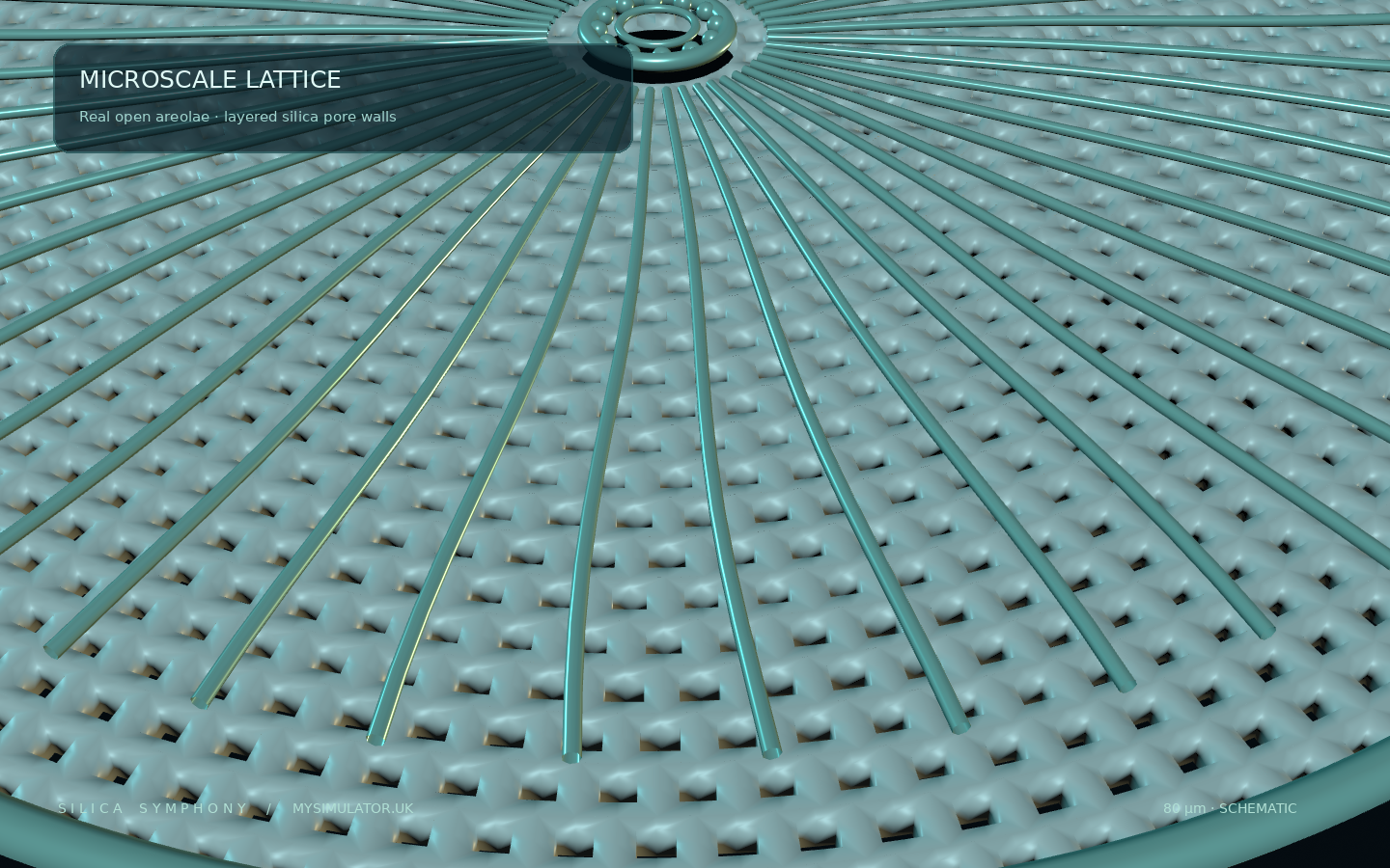
A 3D close-up makes the difference between a printed dot and a real aperture visible. The repeating cells in this illustration are built from procedural polar geometry. Curved radial silica ribs, annular rings and a slightly domed valve surface add readable detail without purporting to represent a named species.
How do the interlocking bands work?
The girdle is made up of one or more silica bands positioned along the side of a valve. Together with the valve, these elements build each theca. In real diatoms the number and form of bands varies, and bands may be added during growth. Using the exploded view slider in the 3D interface separates the upper and lower parts so their arrangement can be seen. The animations are teaching controls, not a simulation of the cell's actual division cycle.
Start with the assembled model. Rotate to an edge view, hide the upper epitheca and then gradually separate the parts. Which bands belong visually to the upper shell and which to the lower? Finally switch on the cutaway to inspect the inside.
What lives inside a silica wall?
Diatoms are living cells, not empty glass sculptures. Photosynthetic structures called chloroplasts convert light energy into chemical energy. In the viewer, a subdued green body and golden structures distinguish the living interior from the harder silica shell. These objects are schematic educational placeholders; their precise number, shape and arrangement are not asserted to reflect a real photographed cell.
The interplay of pores, silica chemistry and optical phenomena has made frustules a subject of active research. Natural microstructured materials may scatter, guide or otherwise modify light. Yet an attractive render cannot tell us measured refractive indices, mechanical strength or biological fitness. Those quantities require suitable experiments, optical modelling and species-specific geometry.
What does the model scale mean?
We assign this fictional object a diameter of 80 micrometres (80 µm), with 1 digital scene unit corresponding to 8 µm. The display is enlarged enormously, as in a digital microscope. This value helps students compare orders of magnitude, but it is not a measurement of a real organism. The viewer's zoom level changes apparent size; any decorative on-screen ruler should not be treated as a calibrated microscope scale bar.
Three model detail levels offer a useful engineering comparison. The hero asset has about 227,000 triangles, while two lighter options preserve the silhouette with progressively fewer pores. Close inspection illustrates how mesh optimisation can balance geometry density, visual readability and performance, especially on mobile devices.
Questions and answers
1. Is this an exact 3D scan of a real diatom?
No. It is an original parametric, biologically informed centric-diatom illustration and is not assigned to a real species. It contains a combination of recognisable structural features and deliberate simplifications.
2. What is the difference between a valve and a frustule?
The frustule is the silica cell wall as a whole. A valve is a major face of one shell half; each theca also includes girdle components.
3. What are areolae?
Areolae are pores or perforations in the mineralised valve wall. Their detailed architecture can contain chambers and smaller openings or porous covering structures.
4. Are all diatoms round?
No. Centric diatoms often show radial symmetry, while pennate diatoms are commonly elongated and bilaterally symmetric; both broad groups are diverse.
5. Is the silica identical to ordinary window glass?
No. Diatoms form biologically controlled hydrated amorphous silica, which differs in composition, formation and internal structure from commercial glass.
6. Why does the model have several detail levels?
They demonstrate geometry-performance trade-offs: removing many small pores greatly reduces the data that must be transferred, decoded and rendered.
7. Why do the shell halves overlap?
The epitheca and hypotheca are often described as box-and-lid elements, with the smaller portion fitting inside the larger. Girdle bands make up much of the sidewall.
8. Can the viewer calculate diatom strength or flow through its pores?
No. The geometry is intended for visual exploration and does not include a calibrated fluid, structural or light-propagation solver. Scientific results would need measurements, boundary conditions and validated material parameters.
Reading and attribution
The 3D asset, textures and screenshots were created for this exercise. The model references scientific descriptions but does not reproduce microscopy images or claim ownership of research materials. For further reading see:
- Diatoms of North America — Coscinodiscus guide (shell morphology and areola examples).
- Frontiers in Plant Science (2022) — Frustule-associated proteins and shell components (valves, thecae and girdle bands).
- Revealing the static and dynamic nanomechanical properties of diatom frustules (hierarchical porosity and material architecture).
- University College London — Diatoms and morphology (micropalaeontology overview).
Continue investigating: Return to the interactive frustule explorer to rotate, separate and compare the shell components.